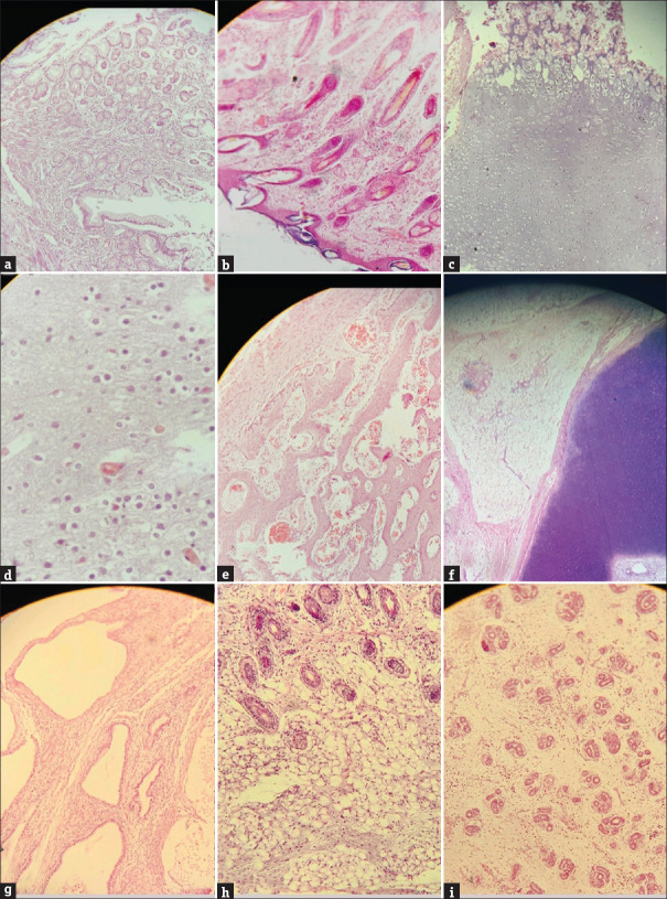
Figure 2.
(a) Glandular epithelium with lamina propria (H and E, ×10), (b) Tissue lined by squamous epithelium with subepithelium showing adnexal structures which are unremarkable (H and E, ×10), (c) Choroid tissue (H and E, ×10), (d) Brain parenchyma with blood vessels (H and E, ×40) (e) Anastomosing bony trabeculae with red blood cells (H and E, ×40), (f) Chondroid tissue on the right side with adipose tissue adjacent to it (H and E, ×10), (g) Cystically dilated glandular cells with fibrous stroma in between (H and E, ×40), (h) Adnexal structures along with adipose tissue (H and E, ×10), (i) Glandular epithelium with loose stoma in between (H and E, ×10)

