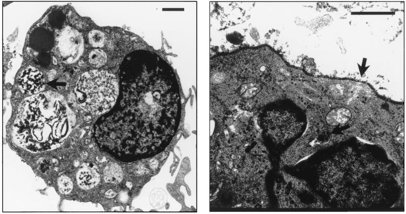
FIG. 3.
Ultrastructural analysis of polyomavirus-infected DC. FACS-sorted CD11c+ DC were infected with polyomavirus (MOI = 3) and prepared for electron microscopy at 36 h postinfection. The length of each inserted line represents 1 μm. Arrows point toward polyomavirus virions within cytoplasmic vacuoles (left panel) and packed along the cellular surface and within a cytoplasmic lamellar structure (right panel). Left panel magnification, ×6,500; right panel magnification, ×43,850.

