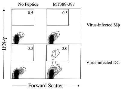
FIG. 6.
Virus-infected DC, but not infected Mφ, prime antipolyomavirus CD8+ T cells in vivo. Uninfected or virus-infected DC and Mφ were injected s.c. in the hind footpads. Six days later, spleen cells were stimulated with MT389-397 for 6 h and then stained for surface CD8 and intracellular IFN-γ. The plots are gated on CD8+ cells, and the values indicate the percentage of cells in the indicated regions. Both axes are log scale.

