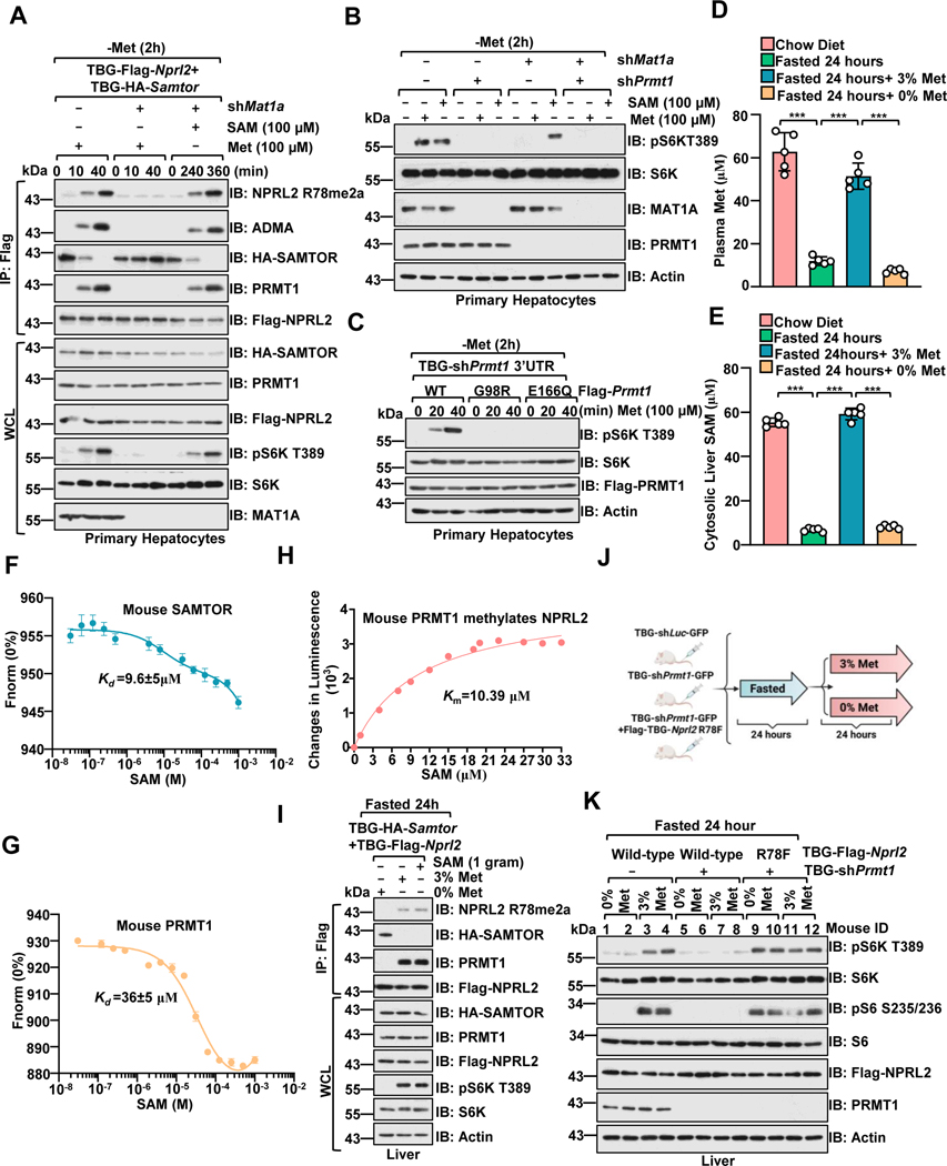
Figure 5. PRMT1 is a physiological methionine/SAM sensor for mTORC1.
A, Primary hepatocytes expressing Flag-Nprl2/HA-Samtor were infected with TBG-shLuc or TBG-shMat1a shRNA. At 72 hours post-infection, cells were deprived of methionine for 2 hour and restimulated with methionine (100 μM) or SAM (100 μM) for the indicated time. The WCL and anti-FLAG IPs were analyzed via immunoblotting with the indicated antibodies.
B, Primary hepatocytes were infected with the indicated shRNA. Cells were then deprived of methionine for 2 hours and restimulated with methionine (100 μM) for 20 mins or SAM (100 μM) for 6 hours. Cell lysates were analyzed by immunoblotting with the indicated antibodies.
C, Primary hepatocytes were infected as indicated. After 72 hours, cells were treated and analyzed as described in A.
D-E, Methionine deprivation significantly reduced SAM levels in the liver. Mice were fasted for 24 hours and refed with 3% or 0% methionine diet for 24 hours. Plasma methionine levels were analyzed via ELISA. (D) Liver tissues were collected for cytosolic SAM level measurement by ELISA (E) (n=6 per group, ***p<0.001, unpaired, two-tailed Student’s t-test).
F-G, The Kd of mouse SAM-SAMTOR and SAM-PRMT1 as determined by the MST assay. n=3 biological repeats were presented.
H, The Km of SAM for mouse PRMT1 to mediate the methylation of NPRL2. Purified NPRL2 proteins (10 ng) were incubated with mouse PRMT1 (20 ng) together with the indicated concentrations of SAM, and the level of SAH generation was analyzed via the MTase-Glo™ Methyltransferase Assay kit. n=3 biological repeats were presented.
I, Dietary methionine regulates Prmt1-Nprl2-Samtor interaction and Prmt1-mediated Nprl2 methylation in the liver. Wild-type male mice with hepatic expression of TBG-Flag-Nprl2 and TBG-HA-Samtor were treated as in D, and the liver lysates were subjected to anti-Flag immunoprecipitation (IP). WCL and IPs were analyzed by immunoblotting with the indicated antibodies.
J-K, A schematic illustration of the experimental setup for studying methionine sensing in vivo. Mice were injected with the respective adenovirus for 21 days to knock down endogenous Prmt1 and overexpress Nprl2 (wild-type or R78F mutant) in the liver as indicated. Mice were then fasted for 24 hours and refed with 3% or 0% methionine diet for 24 hours (J). Liver tissues prepared from the indicated mice were analyzed by immunoblotting with the indicated antibodies (K) (n=2 per group).
See also Figure S5.

