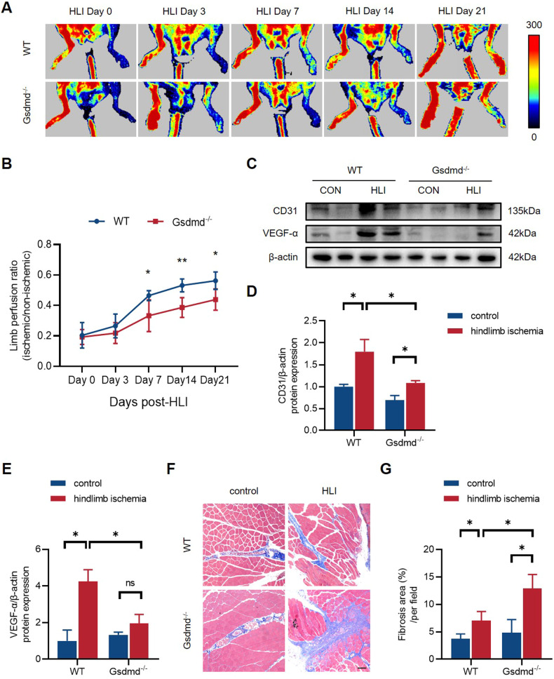
Fig. 3. GSDMD deficiency inhibits perfusion recovery and angiogenesis in vivo after HLI.
A Representative images of limb perfusion in WT and Gsdmd-/- mice analyzed by Laser Doppler Perfusion Imaging and B quantitative Laser Doppler analysis measured by limb perfusion ratio of ischemic to non-ischemic hindlimbs (n = 5 for each group). C Western blots and D, E quantitative analysis of CD31 and VEGF-α expression levels of gastrocnemius tissues in WT and Gsdmd-/- mice at 21st day post-HLI. F Representative images of Masson staining and G quantitative analysis of fibrosis area in gastrocnemius muscles in WT and Gsdmd-/- mice at 21st day post-HLI. Fibrosis area was measured by Image Pro Plus. Scale bar = 100 μm; n = 3 for each group; ns represents p > 0.05, *p < 0.05, **p < 0.01, ***p < 0.001, #p < 0.0001.

