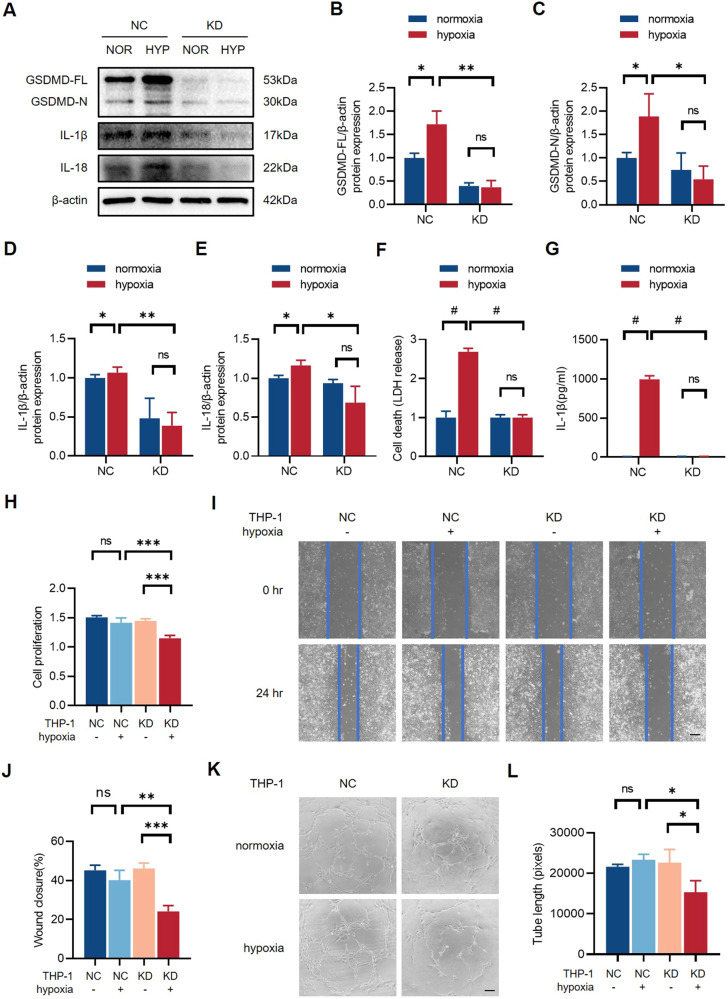
Fig. 5. Inhibition of macrophage pyroptosis damages endothelial cell function.
A Western blots and B–E quantitative analysis of GSDMD-FL, GSDMD-N, IL-1β, and IL-18 expression levels of GSDMD-NC and GSDMD-KD THP-1 cells after normoxia or hypoxia treatment. F The level of LDH in cell culture supernatants detected by LDH release test. G The level of IL-1β in cell culture supernatants detected by ELISA. H Relative cell proliferation rate of HUVECs stimulated with conditioned medium collected from normoxia/hypoxia GSDMD-NC and GSDMD-KD THP-1 cells for 24 h. I, J Representative images and quantification of wound closure for 24 h of HUVECs stimulated with the conditioned medium. K, L Tube formation assay and quantification in HUVECs stimulated with conditioned medium. The values of relative wound closure and tube length were measured by ImageJ. Scale bar = 50 μm. n = 3 for each group; ns represents p > 0.05, *p < 0.05, **p < 0.01, ***p < 0.001, #p < 0.0001.

