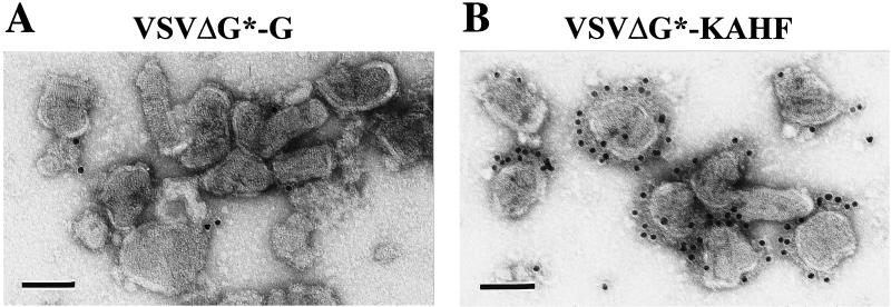
FIG. 3.
Electron microscopy of VSV pseudotype particles. VSVΔG* complemented with the VSV G protein (A) or with the KA H protein and the Edmonston F protein (B) was prepared and partially purified by centrifugation. Viruses were stained with SSPE serum and protein G conjugated with gold, followed by negative staining. Bar, 100 nm.

