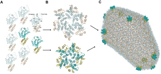
Figure 1.
Assembly of the HIV-1 capsid. (A) Each capsid monomer is shown in cartoon representation, with CA-NTDs in light or dark cyan and CA-CTDs in wheat or olive. Light and dark shades represent monomers that contribute to capsid hexamers and pentamers, respectively. For illustration, on one monomer, α-helices are labelled sequentially, and the positions of N-terminal β-hairpin and Cyp-loop are indicated. (B) Capsid monomers pack into either hexamers (top) or pentamers (bottom). (C) The HIV-1 capsid (PDB: 3J3Q). Approximately 200–250 capsid hexamers combine with 12 capsid pentamers to form a closed fullerene cone structure. Hexamers are shown in cartoon representation. The 12 pentamers are distributed toward the ends of the structure and shown in surface representation.

