Fig. 3.
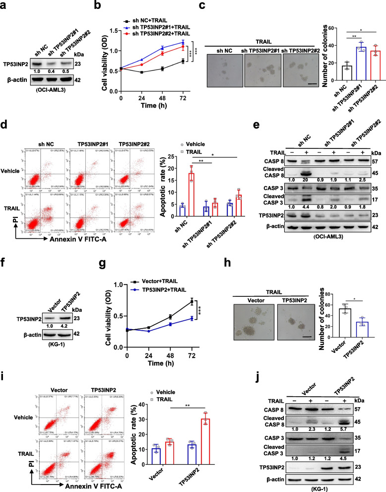
TP53INP2 is a crucial regulator of AML cell response to TRAIL treatment. a Western blot analysis of TP53INP2 in OCI-AML3 cells infected with two sh RNAs targeting TP53INP2. b CCK-8 analysis (n = 3) of cell viability in the OCI-AML3 cells treated with 100 ng/ml TRAIL for 0–72 h. c Colony formation assay was performed in the OCI-AML3 cells (Scale bar: 50 μm). The representative images and quantitative data from three independent experiments were shown in (c, left) and (c, right), respectively. d FCM analysis (d, left) and quantification (d, right) of apoptotic cells in the OCI-AML3 cells. e Western blot analysis of the indicated apoptosis-related proteins in the OCI-AML3 cells. The cells infected with sh NC (scramble shRNA) served as controls in (a-e). f Western blot analysis of TP53INP2 in KG-1 cells transfected with the HA-TP53INP2 plasmids. g CCK-8 analysis (n = 3) of cell viability in the KG-1 cells treated with 100 ng/ml TRAIL for 0–72 h. h Colony formation assay was performed in the KG-1 cells. The representative images and quantitative data from three independent experiments were shown in (h, left) and (h, right), respectively. (Scale bar: 50 μm). i FCM analysis (i, left) and quantification (i, right) of apoptotic cells in the KG-1 cells. j Western blot analysis of the indicated apoptosis-related proteins in the KG-1 cells. The cells transfected with vectors served as controls in (f-j). In (a, e, f, j), β-actin was used as a loading control, and the quantification of protein levels was shown below the protein bands. The data are representative of at least three independent experiments. * p < 0.05, ** p < 0.01, *** p < 0.001

