
Fig. 1.
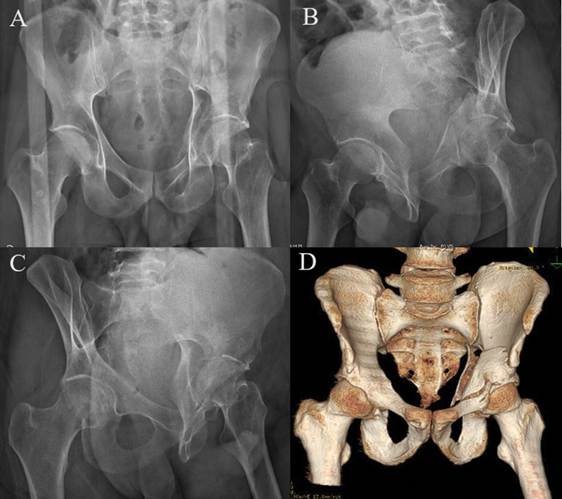
( A ) Anteroposterior (AP) radiograph. ( B ) Obturator oblique radiograph. ( C ) Alar radiograph. ( D ) Three-dimensional (3D) computed tomography showing double column fracture of the left acetabulum.
Official websites use .gov
A
.gov website belongs to an official
government organization in the United States.
Secure .gov websites use HTTPS
A lock (
) or https:// means you've safely
connected to the .gov website. Share sensitive
information only on official, secure websites.
( A ) Anteroposterior (AP) radiograph. ( B ) Obturator oblique radiograph. ( C ) Alar radiograph. ( D ) Three-dimensional (3D) computed tomography showing double column fracture of the left acetabulum.