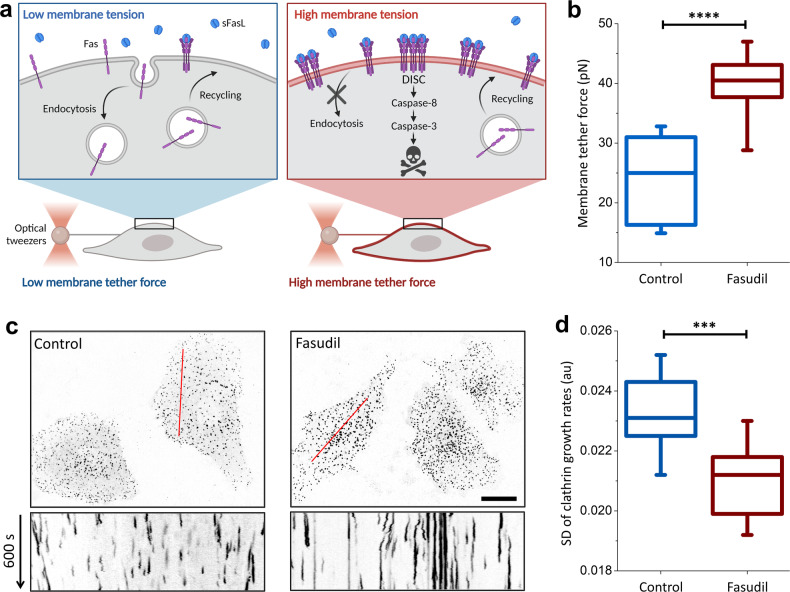
Fig. 1. Mechano-inhibition of endocytosis by fasudil.
a Schematic representation of the study hypothesis: internalization of Fas from the cell surface reduces the sensitivity of cancer cells to Fas-induced apoptosis. Inhibition of endocytosis pathways is expected to increase Fas expression on the cell surface and enhance the formation of the death-inducing signaling complex (DISC) in the presence of sFasL. Endocytosis is slowed by increasing plasma membrane tension, which can be quantified using optical tweezers to measure the membrane tether force. b SUM159 cells treated with 40 µM fasudil showed significantly higher membrane tension than did untreated cells. Ncells = 19 (untreated) and 28 (fasudil). c Endocytic clathrin coats were imaged at the ventral surface of live SUM159 cells (genome edited to express AP2-EGFP) that were untreated (left) or treated with 40 µM fasudil for two hours (right). Kymographs obtained along the marked regions show clathrin-mediated endocytosis dynamics, with short streaks representing fast endocytic events. Streak length increased as endocytosis dynamics slowed. Fluorescence images are inverted to increase visibility. d Standard deviation (SD) of the clathrin growth rates significantly reduced after treatment with 40 µM fasudil. Ncells = 14, Nevents = 84230. ****p < 0.0001, ***p < 0.001; two-tailed t test. Scale bar, 20 µm.

