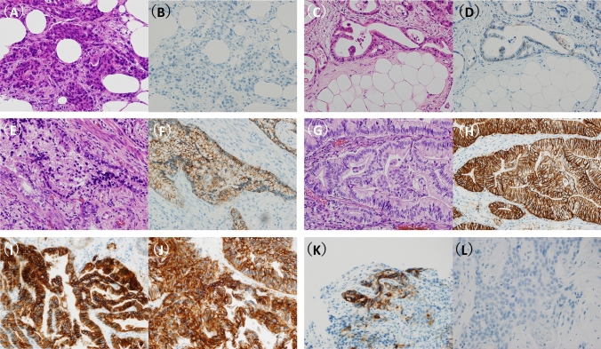
Fig. 2.
Representative images of CLDN18 immunohistochemistry. Pair images (H&E staining and CLDN18 immunohistochemistry) of four categories in CLDN18 intensity: 0, no expression (A and B); 1 + , weak expression (C and D); 2 + , moderate expression (E and F); and 3 + , strong expression (G and H). CLDN18 immunostainings of a representative case with CLDN18-positive in both the primary (I) and peritoneal dissemination (J). CLDN18 immunostainings of a case with discordant results that showed CLDN18-positive in the primary tumor (K) but negative in the peritoneal dissemination (L)

