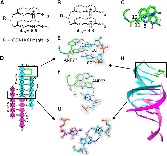
Figure 1.
Interactions of ANP77 with G2C4 RNA. (A, B) chemical structure of the ANP77 molecule: (A) in single and (B) double-protonated state. (C) Stacked conformation of ANP77 observed in the crystal structure. Selected atoms are indicated (see text for details). (D) secondary structure of G2C4–ANP77 complex. Green lines represent ANP77 ligand. (E, F) Pseudo-canonical base-pairs formed between ANP77 (green) and cytosine residues from chain A (cyan). (G) Base triple between cytosine from chain C (purple), guanosine from chain A (cyan), and cytosine from chain B (cyan). (H) Crystal model of RNA tetramer (chain A and B in cyan; chain C and D in pink) with bound ligand molecule (green sticks). The 2Fo–Fc electron density map (gray) is contoured at the 1σ level. The H-bonds are represented by black dashed lines.

