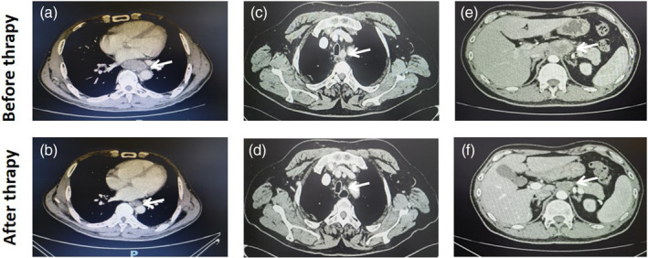
FIGURE 2.

The representative enhanced computed tomography images before and after neoadjuvant chemotherapy and immunotherapy. (a, b) Case 1 had cancer in the lower esophagus with cT3 stage disease before therapy, and pathological diagnosis subsequently confirmed no residual cancer (ypT0). (c, d) Case 2 had lymph node metastasis around the left recurrent laryngeal nerve (cN+), and pathological diagnosis confirmed the node to be metastatic (ypN+). (e, f) Case 3 had bulky lymph node metastasis around the gastric left artery (cN+), and pathological diagnosis confirmed no residual cancer in the nodes (ypN−).
