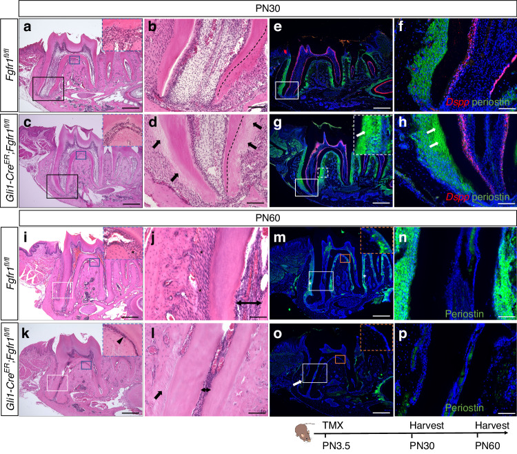
Fig. 3.
Narrowed PDL space with ankylosed tooth root in Gli1-CreER;Fgfr1fl/fl mice. a–d Histological analysis of Fgfr1fl/fl and Gli1-CreER;Fgfr1fl/fl mice at PN30. Black arrows point to abnormal cementum. Black dashed lines outline the interface between root dentin and cementum. e–h Expression of Dspp and periostin in Fgfr1fl/fl and Gli1-CreER;Fgfr1fl/fl mice. White arrows point to abnormal periodontal ligaments. i–l Histological analysis of Fgfr1fl/fl and Gli1-CreER;Fgfr1fl/fl mice at PN60. The black asterisk points to periodontal ligament space; the black arrowhead points to narrowed periodontal ligament space in furcation; the black arrow points to the absence of periodontal ligament space where the tooth root connects to alveolar bone; Line with arrows indicates root pulp cavity. m–p Periostin expression in Fgfr1fl/fl and Gli1-CreER;Fgfr1fl/fl mice at PN60. The white asterisk points to the periodontal ligament; the white arrow points to the absence of periostin expression. The Schematic at the bottom indicates the induction protocol. Scale bars, b, d, f, h, j, l, n, p, 100 μm; others, 500 μm

