Fig. 8.
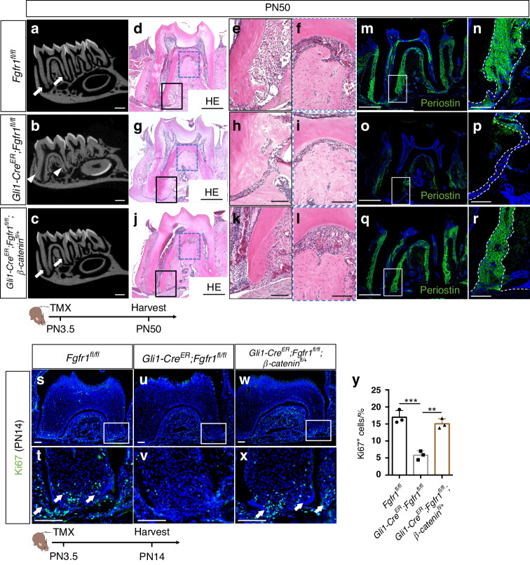
Downregulation of WNT signaling rescues tooth ankylosis in Gli1-CreER;Fgfr1fl/fl mice. a–c MicroCT analysis of the first mandibular molars in Fgfr1fl/fl, Gli1-CreER;Fgfr1fl/fl and Gli1-CreER;Fgfr1fl/fl;β-cateninfl/+ mice at PN50. White arrows point to the periodontal ligament space; white arrowheads point to the narrowed periodontal ligament space. d–l Histological analysis of Fgfr1fl/fl, Gli1-CreER;Fgfr1fl/fl and Gli1-CreER;Fgfr1fl/fl;β-cateninfl/+ mice. m–r Periostin expression in Fgfr1fl/fl, Gli1-CreER;Fgfr1fl/fl and Gli1-CreER;Fgfr1fl/fl;β-cateninfl/+ mice. Space between white dashed lines (n, p, and r) indicates periodontal ligament space. s–x Proliferation stained with Ki67 in Fgfr1fl/fl, Gli1-CreER;Fgfr1fl/fl and Gli1-CreER;Fgfr1fl/fl;β-cateninfl/+ mice. White arrows point to Ki67+ cells. y Quantification of Ki67+ cells in three groups. Fgfr1fl/fl versus Gli1-CreER;Fgfr1fl/fl: P = 0.000 4; Gli1-CreER;Fgfr1fl/fl versus Gli1-CreER;Fgfr1fl/fl;β-cateninfl/+: P = 0.001, n = 3 biologically independent samples, with one-way ANOVA performed. The Schematic at the bottom indicates the induction protocol. **P < 0.01, ***P < 0.001. Scale bars, a–c, 1 mm; d, g, j, m, o, q, 500 μm; others, 100 μm

