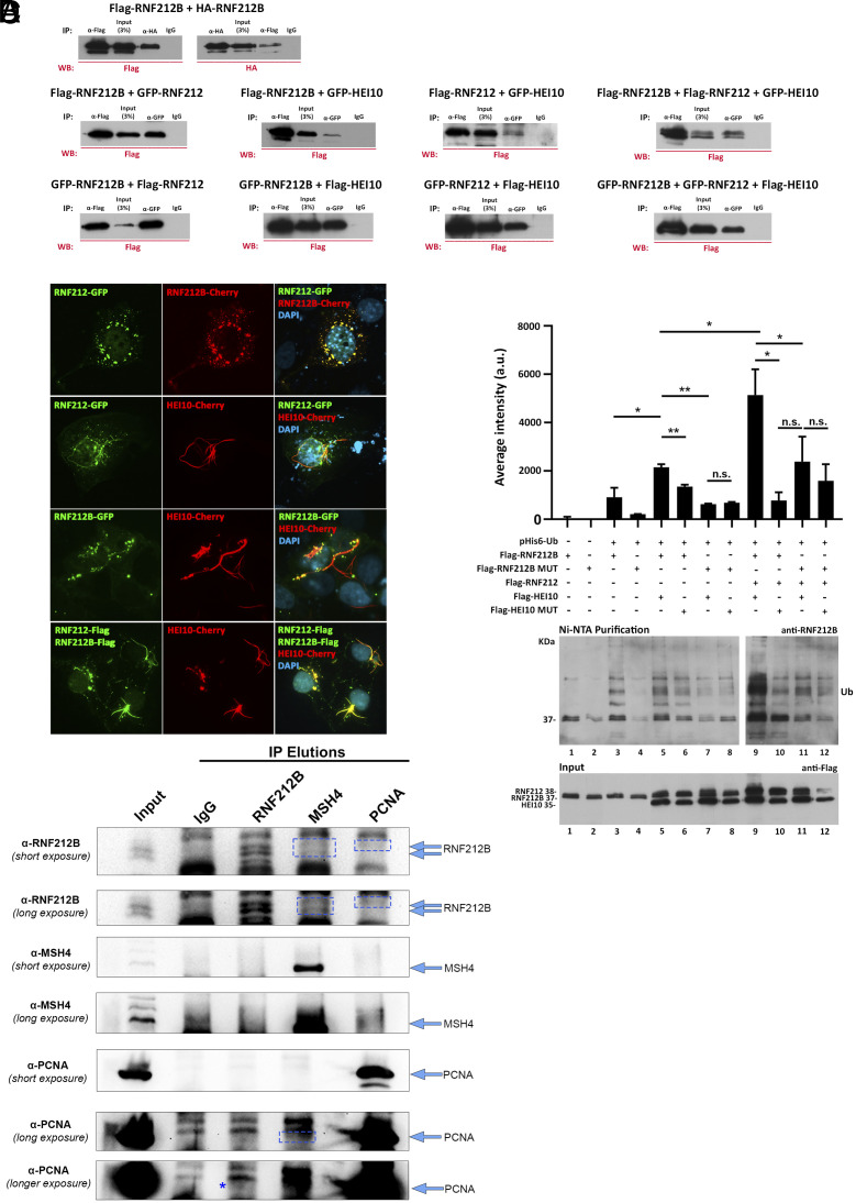
Fig. 3.
RNF212B interactions and Ubiquitylation activity. (A) Co immunoprecipitation of RNF212B with RNF212B, RNF212, and HEI10. HEK293T cells were cotransfected with plasmids encoding RNF212, RNF212B, and HEI10 tagged with FLAG or GFP. Protein complexes were immunoprecipitated overnight with an antibody anti-FLAG, anti-GFP, or IgGs (as a negative control) and analyzed by immunoblotting. Positive interaction of RNF212B with RNF212 and HEI10 as well as RNF212B (self-interaction) is shown. Additionally, a positive interaction between RNF212 and HEI10 is observed. (B) COS7 cells immunofluorescence shows colocalization of transfected RNF212B with RNF212 and HEI10. COS7 cells were transfected to express RNF212B (fused to either Cherry, Flag, or GFP) along with RNF212 (fused with Flag or GFP) and/or HEI10 (Cherry-tagged). Nuclear and punctuate cytoplasmic RNF212B signal perfectly colocalizes with RNF212 signal (Top). Transfected HEI10 forms a ring fiber-like pattern that when cotransfected with RNF212 and/or RNF212B recruits them to this ring structures altering their localization pattern. (Scale bars: 10 μm.) (C) RNF212B has autoubiquitynilation activity. HEK293T cells were cotransfected with Flag-tagged plasmids encoding RNF212, RNF212B (WT or mutant), and/or HEI10 (WT or mutant) in the presence or absence of a plasmid encoding 6xHis-Ubiquitin. Protein complexes were purified using Ni-NTA, favoring the purification of proteins containing 6xHis sequence. Then, ubiquitylation was analyzed by immunoblotting using antibodies anti-RNF212B and anti-Flag (Input). The results show RNF212B autoubiquitinylation. As observed in the statistics of the quantification of average intensity for each well, the strongest ubiquitinylation was exhibited when RNF212B was cotransfected along with RNF212 and HEI10 and was abolished when a RNF212B dead mutant was transfected. Quantification of the results of three independent experiments. Two-tailed Welch’s t test analysis: *P < 0.05. **P < 0.005. (D) IPs in isolated germ cells with the antibodies indicated (Top) against RNF212B, MSH4, and PCNA, and western blot analysis of the eluted proteins with the same antibodies (Left) detecting specific bands for RNF212B in the MSH4 and PCNA elution, and PCNA in the RNF212B and MSH4 elution (boxes and asterisk). Arrows indicate sizes for bands of interest in each blot.

