Fig. 1.
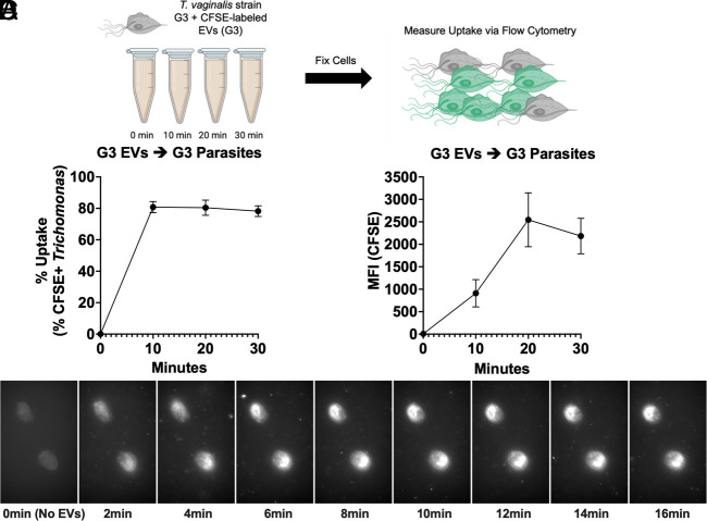
Trichomonas-derived EVs are taken up by T. vaginalis. (A) Schematic of TvEV uptake assay. T. vaginalis strain G3 was exposed to 10 µg/mL of CFSE-labeled G3-EVs for 0, 10, 20, and 30 min and TvEV uptake was measured via flow cytometry. Created with BioRender.com. (B and C) Percent uptake and MFI of CFSE-positive parasites plotted over time. Percent uptake was measured by the percent of CFSE-positive parasites in the entire population and the amount of TvEV uptake was quantified by the increase in MFI plotted over time. Dots = mean ± SD. N = 3 replicates/experiment, three experiments total. (D) Individual images from 2-min intervals of time-lapse video showing uptake of ExoGlowTM labeled G3-EVs by T. vaginalis.

