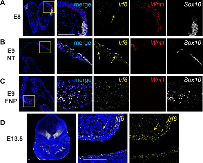
Fig. 1.
Irf6 is expressed with neural crest cell markers Wnt1 and Sox10 in neural folds and neural tube during early embryogenesis. In situ hybridization of Irf6 (yellow), Wnt1 (red), and Sox10 (white) RNA transcripts. A. Coronal section of E8 mouse embryo (dorsal to top) showing the neural fold. In situ hybridization shows RNA expression domains of Irf6, Wnt1, and Sox10, where Irf6 and Wnt1 transcripts are found in the same regions of the neural tube, highlighted by yellow arrow. Box indicates area of higher magnification to the right. B. Sagittal section of E9 mouse embryo (cranial to left). Box indicates a magnified portion of the neural tube. Irf6 is expressed in the neuroectoderm and overlaps with Wnt1 and Sox10 expression (yellow arrows). C. Sagittal section of E9 mouse embryo (cranial to left). Box indicates a magnified portion of frontonasal prominence (FNP). Irf6 is expressed in the FNP mesenchyme, along with the migratory NCC marker Sox10. D. Coronal section of E13.5 embryo (dorsal to top). Box indicates higher magnification of palate shelf epithelium and mesenchyme. Irf6 is highly expressed in the basal epithelium and periderm and the palate mesenchyme (yellow arrow). Blue is dapi. Scale: 100 uM.

