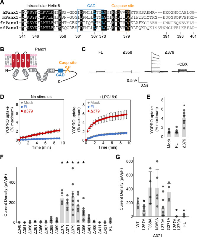
Figure 1. Systematic truncation uncovers a C-terminal activating domain.
(A) Sequence alignment of the listed Panx1 orthologues around the CAD. (B) A schematic representation of the full length Panx1. IH stands for internal helix. (C) Exemplar whole cell patch clamp recordings of the wild type and C-terminally truncated Panx1 constructs. Cells were clamped at −70 mV and stepped from −110 mV to +110 mV for 0.5 s in 20 mV increments. CBX was applied at 100 μM. (C) and (D) YO-PRO-1 uptake triggered by LPC16:0 (10 μM). Averaged YO-PRO-1 uptake (D) and comparison of the signals at 5 min (E) are shown. N=7–10 and error bars indicate SEM. (F) and (G) Peak whole-cell current density at +110mV obtained from the Panx1 truncation constructs (F) and alanine substitutions of Panx1Δ371 (G). N=4–16 and error bars indicate SEM. Asterisks indicate significance of p<0.05 determined by one-way ANOVA followed by Dunnett’s test comparing the full length Panx1 (FL) to each construct.

