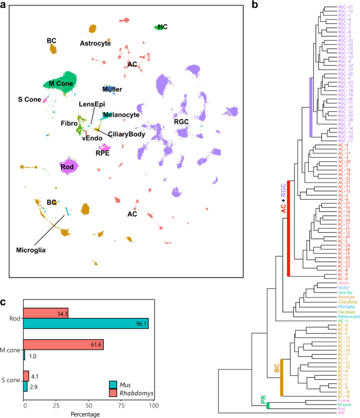
Figure 5. A retinal cell atlas for Rhabdomys.
a) Transcriptionally distinct clusters of Rhabdomys retinal cells visualized using UMAP58. Cells are coloured by class identity (RPE, retinal pigment epithelial cells; vEndo, vascular endothelial cells; Lens Epi, lens epithelial cells; Fibro, fibroblasts). b) Dendogram showing transcriptional relationships of Rhabdomys cell clusters, with major clades corresponding to cell classes. c) Bar chart indicating proportion of photoreceptor types in Rhabdomys (red) and Mus (cyan).

