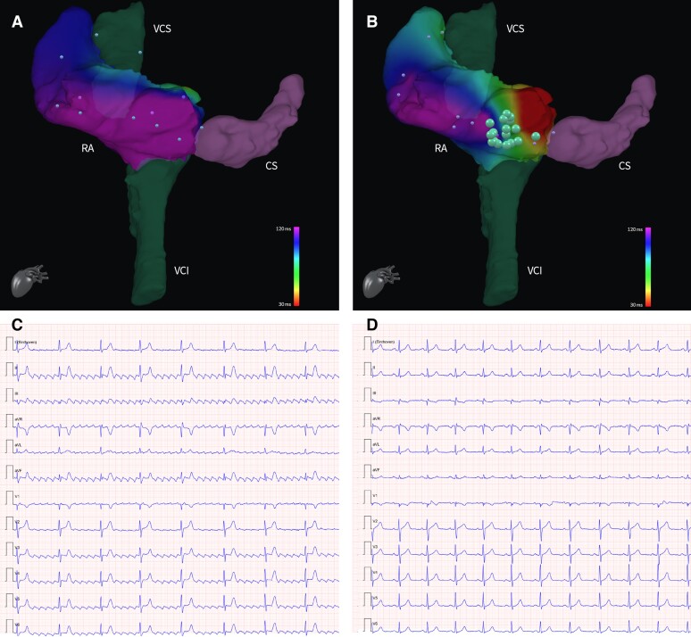
Figure 2.
Data of mapping points (little dots) derived from the Advantage-MR EP Recorder/Stimulator System can be used to create activation maps to superimpose on the 3D anatomical shell. Panel (A) shows the activation map of the RA of our patient at the beginning of the procedure, corresponding to a typical counterclockwise atrial flutter (C). After the ablation, the new activation map reflects the achievement of a bidirectional bock at the level of the isthmus (B), with achievement of sinus rhythm (D). Ablation points (big dots) can interactively show information as power, duration, impedance drop, and temperature associated to the RF delivery. 3D, three-dimensional; CS, coronary sinus; IVC, inferior vena cava; RA, right atrium; RF, radiofrequency; SVC, superior vena cava.

