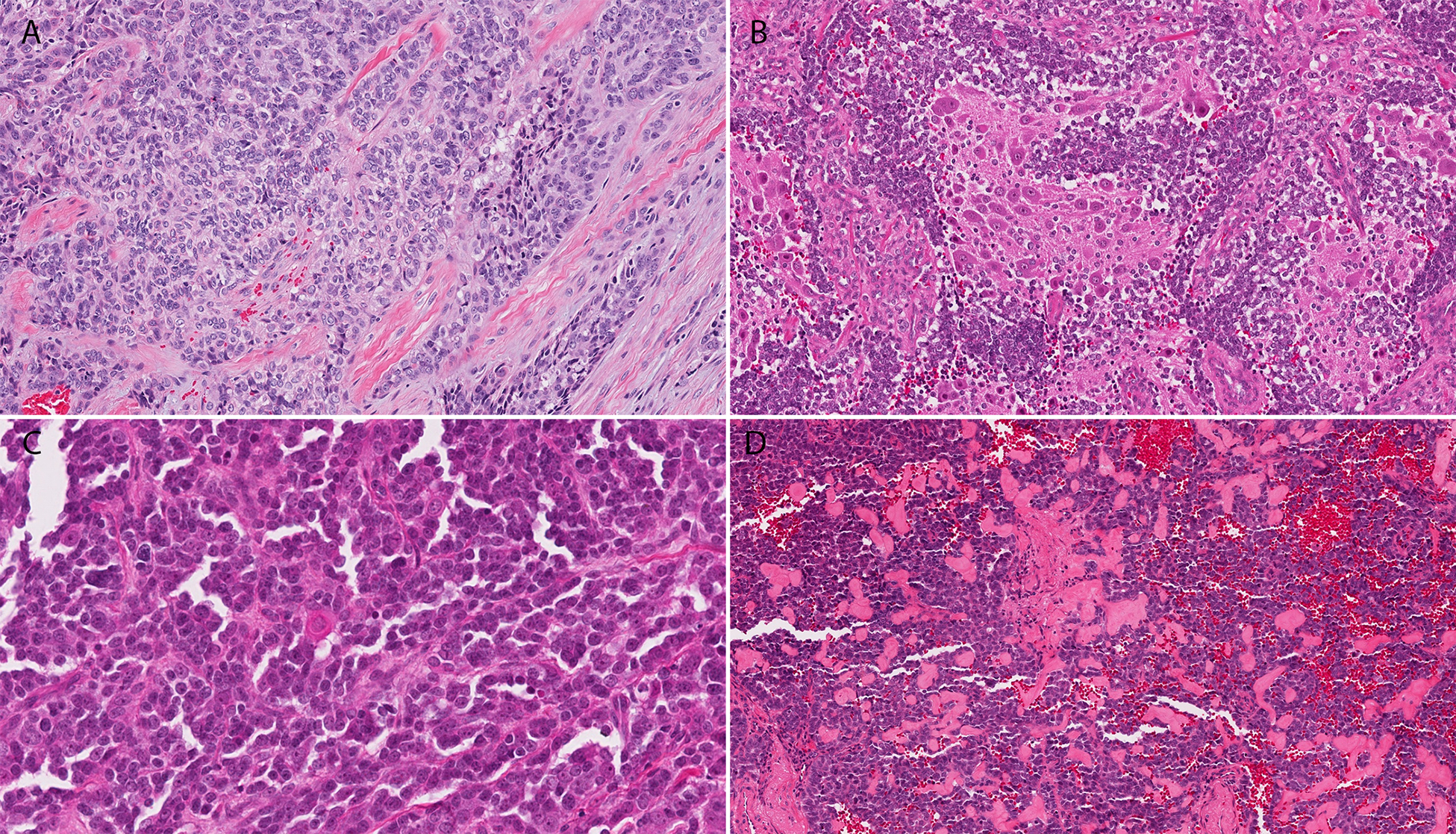
Figure 2.

The cytoplasm of the gingival ALES was more abundant and pale pink (A), while the case which was reviewed post-chemotherapy showed large ganglion-like cells consistent with neuronal maturation (B). Case 4 showed single cell keratinization (C), while case 1 contained deposition of eosinophilic matrix between tumor nests (D).
