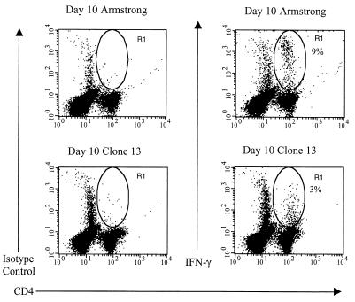
FIG. 4.
Intracellular IFN-γ expression of LCMV peptide-specific CD4+ T cells from LCMV clone 13-infected C57BL/6 mice. Splenocytes from LCMV Armstrong- or clone 13-infected mice (both day 10) were stimulated with the LCMV MHC class II-restricted peptide GP61-80 in the presence of IL-2 and brefeldin A for 5 h and subsequently stained as described in the legend to Fig. 1. The percentages shown each indicate the number of CD4+ T cells that stained positive for intracellular IFN-γ as represented by the R1 gate. Data are representative of three separate experiments, with three mice per experiment.

