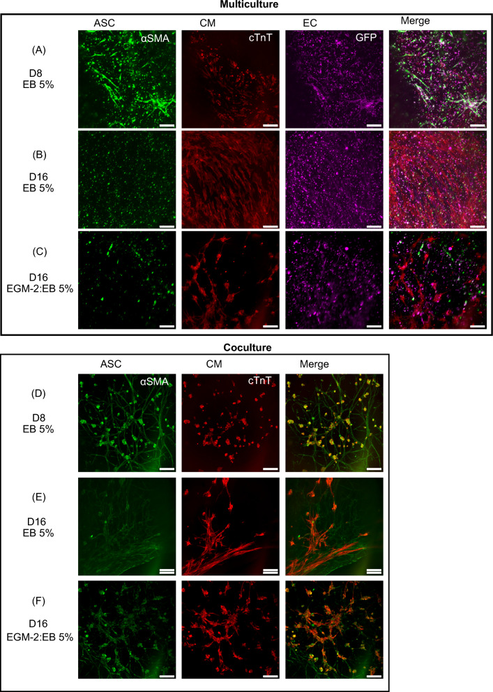
Fig. 4.
Formation of A–C cardiovascular multiculture comprising of human adipose stem/stromal cells (ASC), induced pluripotent stem cell-derived cardiomyocytes (iPSC-CM) and Human Umbilical Vein Endothelial Cells (HUVEC) at day 8 in EB 5% and at d16 in EB5%:EGM-2 and in EB 5%; D–F myocardial co-culture comprising of ASC and iPSC-CM in EB 5% (d8) and in EB 5%:EGM-2 and EB5% (d16) medium. ASC organize into smooth muscle cell-like network from d8 onwards as shown with α-smooth muscle actin staining (D–F). iPSC-CM shift into elongated morphology in both cardiac models as shown with α-troponin t staining (A–F). Vascular network formed by GFP signaling HUVEC degrades from d8 to d16 in multicellular construct (A–C). Scale bar 200 µm

