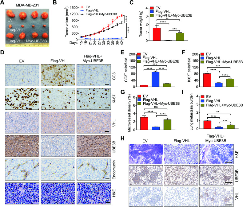
Fig. 4. UBE3B alleviates VHL’s inhibitory effect on breast tumor growth and metastasis.
A–C Tumor images (A), tumor growth curves (B), and tumor weight (C) of mice with orthotopic implantation of EV (empty vector), Flag-VHL, and Flag-VHL + Myc-UBE3B MDA-MB-231 cells into the mammary fat pad (mean ± SD, n = 4). ****P < 0.0001, by 2-way ANOVA Tukey’s multiple comparisons test (B) or 1-way ANOVA Tukey’s multiple comparisons test (C). D–G Representative H&E staining and immunohistochemical staining of Ki-67, CC3 (cleaved caspase-3), and endomucin in primary tumors (D). Scale bars: 100 μm. Ki-67-positive cell numbers (E), CC3-positive cell numbers (F), and microvessel density (G) were quantified. (mean ± SD, n = 4). ***P < 0.001, ****P < 0.0001, by 1-way ANOVA Dunnett’s multiple comparisons test. H, I Representative images of metastases and protein levels of UBE3B and VHL in the lungs analyzed by H&E staining and IHC staining (H), respectively. Lung metastases were quantified by qPCR (H) (mean ± SD, n = 4). **P < 0.01, ***P < 0.001, ****P < 0.0001, by 1-way ANOVA Tukey’s multiple comparisons test. Scale bars: 500 μm.

