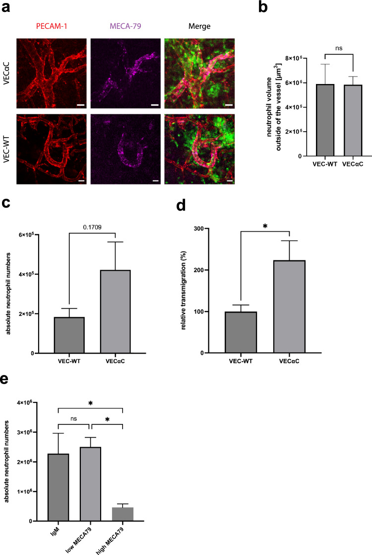
Figure 4.
VEC-αC does not inhibit PMN extravasation through omental HEVs, but increases extravasation through larger, non-HEV venules. (a) Confocal microscopic images of HEVs in the omentum of VEC-WT and VEC-αC mice. Endothelial junctions are stained for PECAM-1 (red), high endothelial venules (HEV) in milky spots by MECA79 (magenta) and neutrophils are genetically labeled by LysM-EGFP (green). Bar = 20 µm. (b) Quantification of the volume occupied by neutrophils outside of HEVs 3 h after IL-1β stimulation in VEC-WT or VEc-αC mice. A total of 29 vessels were evaluated (n = 5 mice VEC-WT, n = 4 mice VEc-αC). P = 0.9717, t-test. (c and d) Absolute numbers (c) and relative numbers (d) of neutrophils isolated from the peritoneal lavage of VEC-WT or VEC-αC mice treated with 50 µg anti-MECA79 antibody to inhibit leukocyte extravasation through HEVs. 3 h after i.p. injection of 50 ng IL-1β peritoneal exudates were collected. Data were analyzed from 3 different experiments, with 9–12 mice in total and are presented as mean ± SEM. * P = 0.0396; t-test. (c and d) are based on the same set of results. (e) Number of neutrophils isolated from the peritoneal lavage of C57Bl/6 mice treated with 3 µg (non-blocking dose, used for staining) or 50 µg anti-MECA79 antibody (blocking dose, inhibition of leukocyte extravasation through HEVs). 3 h after i.p. injection of 50 ng IL-1 peritoneal exudates were collected. Data were analyzed from 9 to 10 mice per group (28 mice in total) and are presented as mean ± SEM. * P ≤ 0.05; one-way ANOVA.

