Figure 7.

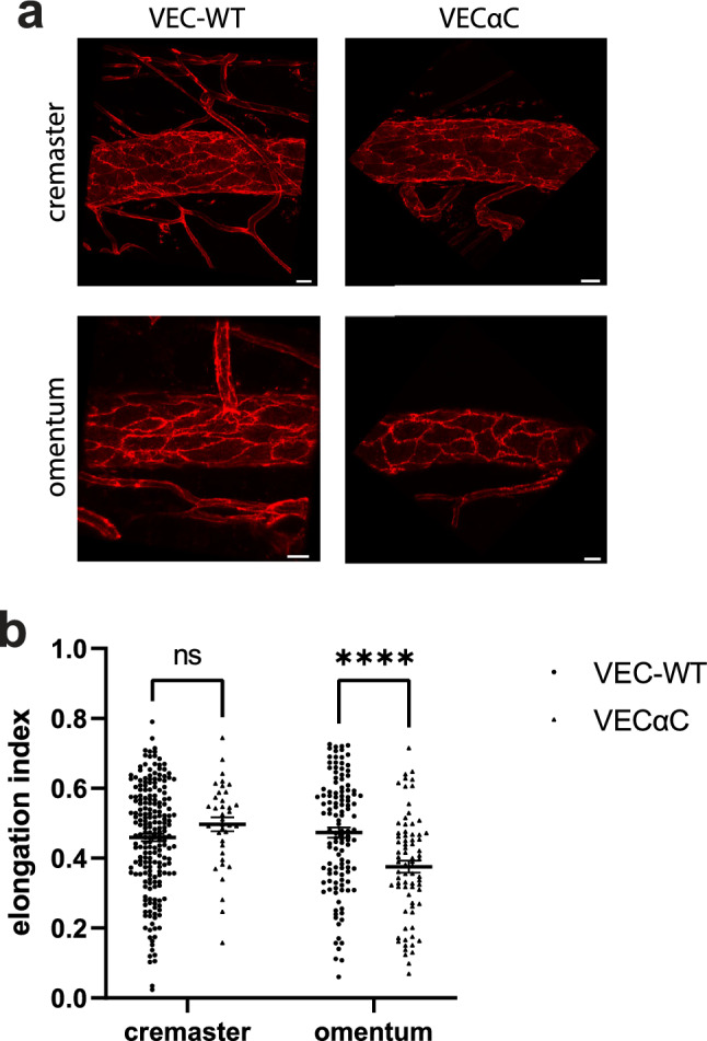
Endothelial cells are less elongated in venules of the omentum of VEC-αC mice. (a) Images of venule segments stained for PECAM-1 from cremaster muscle and omentum from VEC-WT and VEC-αC mice, bar = 20 µm. (b) Quantification of the elongation index of endothelial cells in venules (diameter 60–85 µm) of the cremaster and omentum of VEC-WT and VEC-αC mice. A total of 56 vessels (n = 20 VEC-WT cremaster, n = 17 VEC-αC cremaster, n = 11 VEC-WT omentum, n = 8 VEC-αC omentum) were evaluated and presented as mean ± SEM. ****P < 0.0001; two-way ANOVA.
