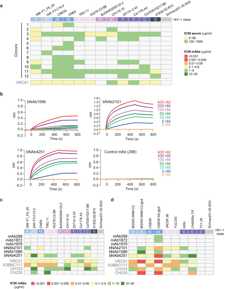
Fig. 5. HIV Env binding and neutralization assays of serum and IgG samples.
a Neutralization assays were performed against 12 viruses from clades A, AC, AE, B, BC, C, and G of tiers 2. The colors of the heatmap correspond to the IC50 of the sera in micrograms per ml. The SIVmac251.30.SG3 virus is used as a negative control. b Antibody–SOSIP interactions were determined by biolayer interferometry (BLI). The mAbs or bNAbs were loaded on a protein A biosensor, dipped into a solution of the SOSIP trimer at different concentrations (ranging from 5 to 400 nM), and the nm shift was recorded. BLI sensorgrams are representative examples of experiments repeated two times (n > 2). c, d Neutralization assays were performed against twelve viruses from clades A, AC, AE, B, BC, C, and G of tiers 2. c The colors of the heatmap correspond to the IC50 in micrograms per ml, for each antibody. The SIVmac251.30.SG3 virus is used as a negative control. d Neutralization assays were performed against glycan-mutated viruses to support epitope mapping to the CD4-binding site. Neutralization assay experiments were repeated two times (n > 2). Source data are provided as a Source Data file.

