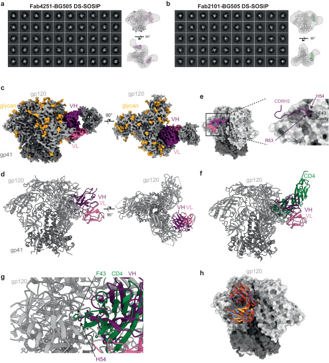
Fig. 6. Fab4251 and Fab2101 interaction with BG505 DS-SOSIP.
a 3D reconstruction of Fab4251-SOSIP complex by nsEM. b 3D reconstruction of Fab2101-SOSIP complex by nsEM. c Side and top views of the cryo-EM density map of the Fab4251-DS-SOSIP complex, with gp120 in light gray, gp41 in dark gray, VH in violet, and VL in pink. d Atomic model of Fab4251-DS-SOSIP complex shown in cartoon representation. e Footprint representation of the heavy and light-chain binding surface on DS-SOSIP, colored according to (c). Inlet on the right represents the CDRH2 loop in violet, with H54 in the F43 cavity. f Overlay of CD4 receptor (green) bound to SOSIP (PDB.5U1F) and Fab4251 (violet). g Close view of VH H54 from Fab4251 and F43 in CD4. h Overlay of VRC01-class antibodies on SOSIP with Fab4251 (violet), VRC01 (PDB.6V8X, green), PG04 (PDB.4I3S, red), and 3BNC60 (PDB.4GW4, orange).

