
Fig 3—
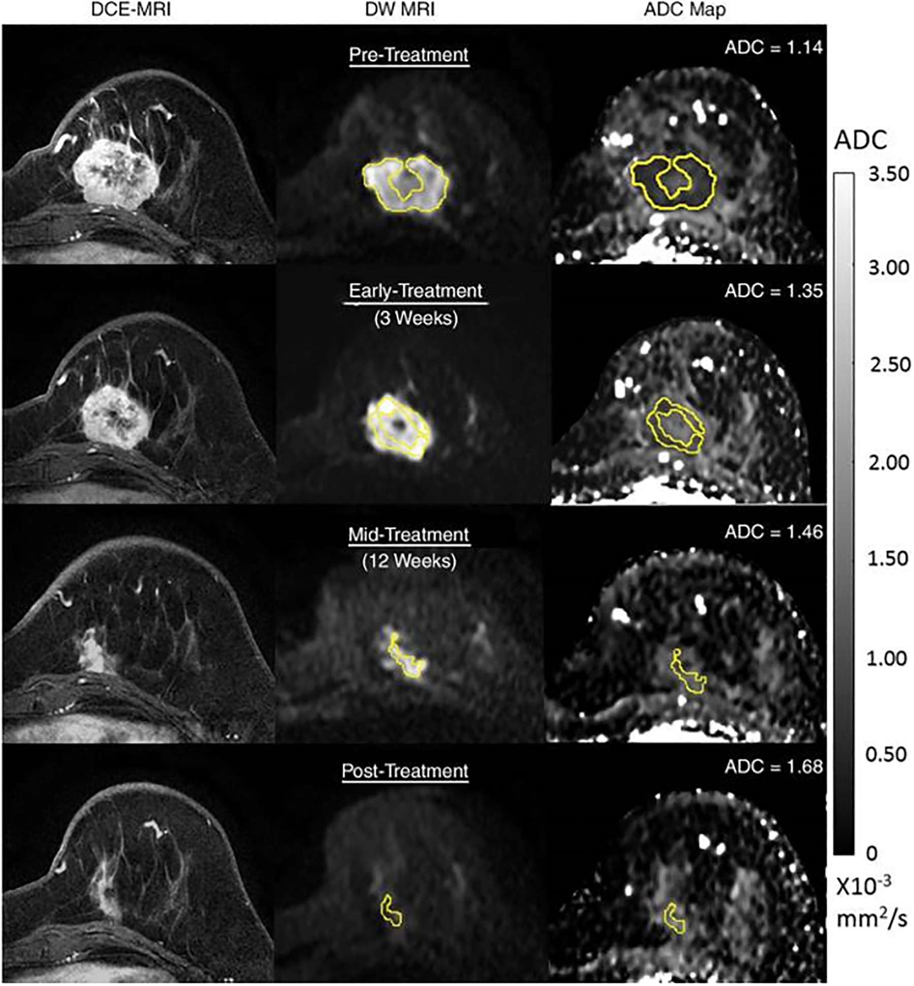
54-year-old woman with grade III triple-negative (hormone receptor–negative/human epidermal growth factor receptor 2–negative) breast cancer. Patient underwent neoadjuvant therapy. Serial 3-T MRI examinations were performed over course of treatment. DCE MRI (left), DWI (b = 800 sec/mm2) (middle), and ADC map (right) before treatment (top row), during early treatment (after 3 weeks) (second row), during midtreatment (after 12 weeks) (third row), and after treatment (bottom row). Tumor measured 4.2 cm before treatment. At each time point, whole-tumor ROI was placed on multiple slices on DWI, avoiding central necrotic area, and mean ADC was calculated for all voxels in 3D ROI. ADC values increased progressively during treatment, yielding ΔADC = 18% at early treatment, 28% at midtreatment, and 47% at posttreatment. After treatment, patient was found to have had pathologic complete response. Reprinted with permission from: Partridge SC, Zhang Z, Newitt DC, et al. Diffusion-weighted MRI Findings Predict Pathologic Response in Neoadjuvant Treatment of Breast Cancer: The ACRIN 6698 Multicenter Trial. Radiology 2018; 289:618–627.