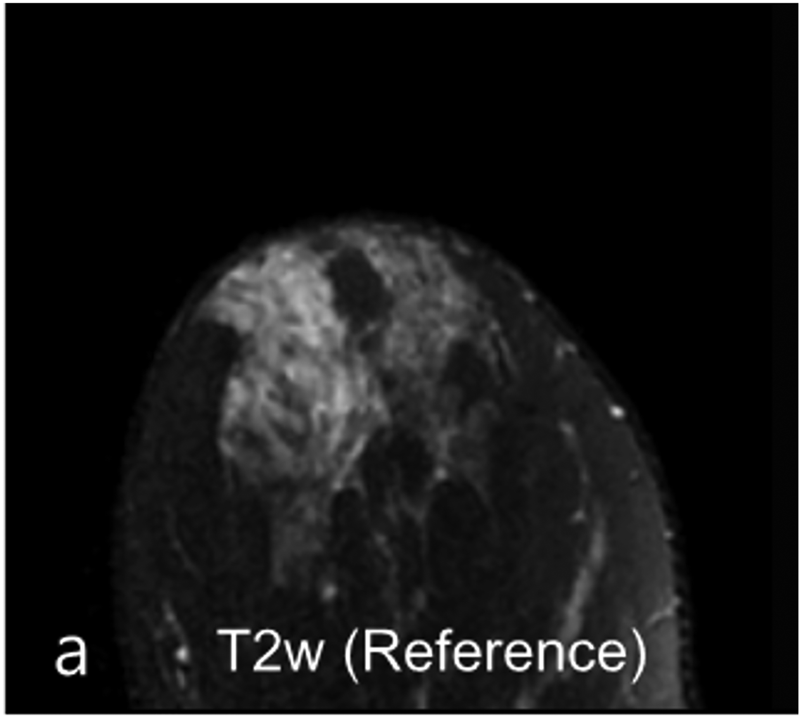
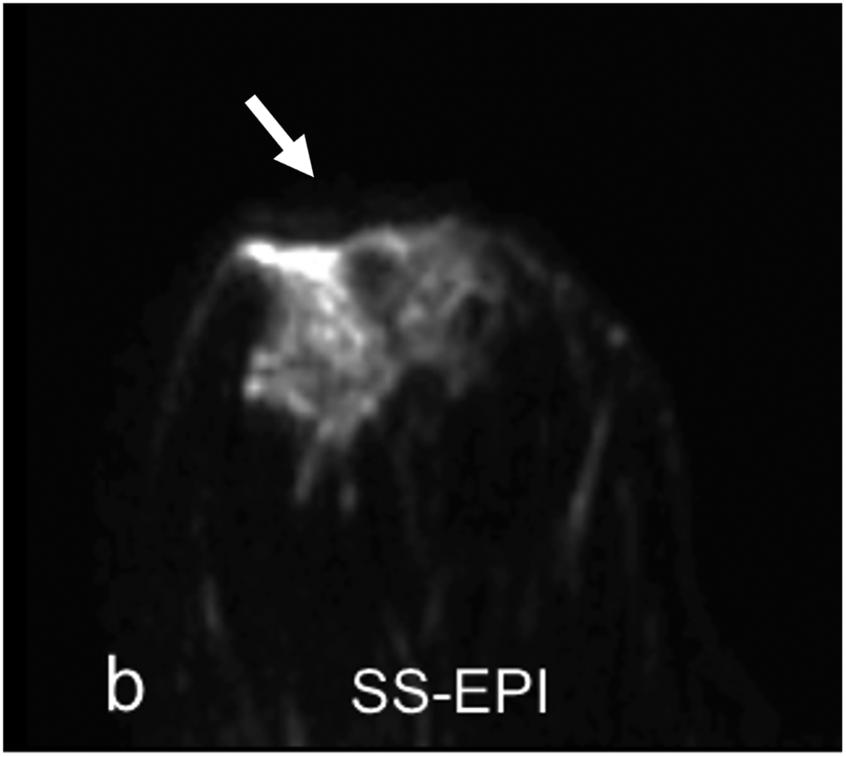
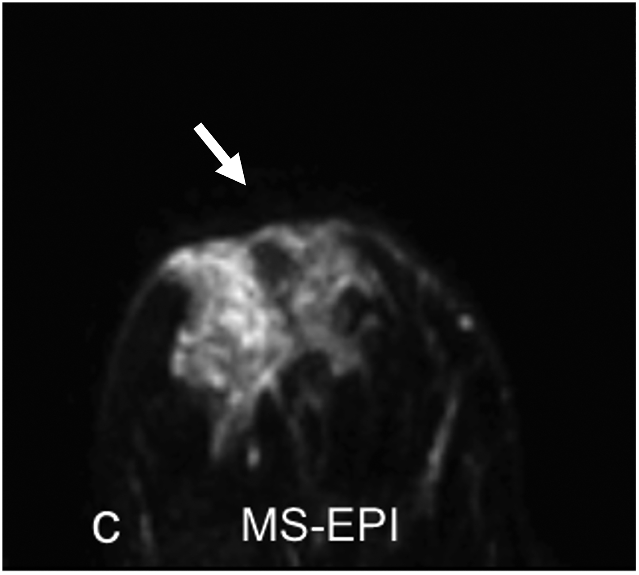
Fig 5—


33-year-old woman undergoing high-risk screening by breast MRI. (a) T2-weighted fast spin-echo image (spatial resolution of 0.8 × 0. 8× 1.3 mm3) serves as anatomic reference. (b-c) DWI with b = 0 s/mm2 obtained using conventional single-shot echo-planar imaging (SS-EPI) (b), and using multi-shot EPI (c) (MS-EPI; Philips Healthcare IRIS technique, 2 shots), both with high spatial resolution of 1.2 × 1.2 × 4 mm3. Distortions related to magnetic susceptibility effects in anterior breast (arrow, b and c) are reduced using MS-EPI acquisition compared to SS-EPI acquisition. Examination shows no suspicious breast findings.
