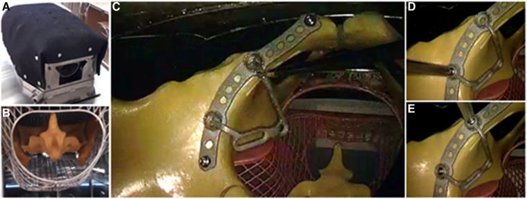
Figure 2.
Experimental setup: (A) The laparoscopic training torso in which the SYNBONE pelvis is located. (B) View from below into the training torso, the pelvis with sacrum can be seen. (C) View via endoscope from cranial into the pelvis lying in the training torso. The buttress part of the plate is being connected to the baseplate part that has already been inserted and is fixed (D) with a dorsal and (E) ventral screw.

