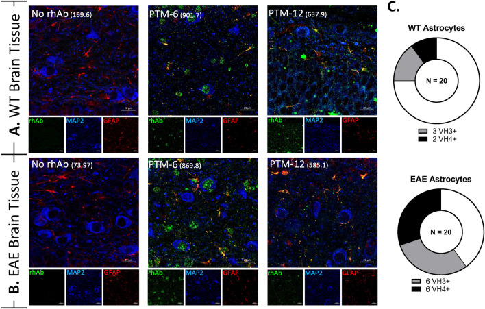
Fig. 4.
Plasmablast rhAbs bind neurons and astrocytes in mouse brain. A, B Representative images of rhAbs binding neurons and astrocytes in the brain of WT (A) and EAE (B) mice. Green: IgG staining. Red: GFAP. Violet: MAP2. Blue: DAPI. In the merge panels, yellow indicates co-stain of GFAP with the rhAb. The MFI of astrocytic staining is indicated in parentheses. Scale bar: 20 μm. C Pie charts summarizing the number of rhAbs binding astrocytes in WT and EAE spinal cord tissue. Positive binding rhAbs are binned according to antibody variable heavy chain use (VH3+ or VH4+)

