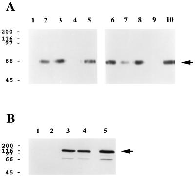
FIG. 3.
HHV-7 gp65 exhibits heparin-binding activity. (A) Insect cell culture supernatants containing soluble recombinant gp65-(His6) were incubated with heparin-acrylate beads in the absence (lanes 5 and 10; negative control) or presence of an excess amount of various glycosaminoglycans, including heparin (lanes 4 and 9; positive control), heparan sulfate (lane 1), N-acetylheparin (lane 2), de-N-sulfated heparin (lane 3), and the chondroitin sulfates A, B, and C (lanes 6, 7, and 8, respectively). After a washing, the beads were boiled in SDS-PAGE sample buffer, and eluted proteins were separated by electrophoresis on an SDS–12.5% polyacrylamide gel and transferred to nitrocellulose. Immunoblot analysis was then conducted using an anti-His6 monoclonal antibody and a chemiluminescent detection system (see Materials and Methods). (B) Lysates from COS-1 cells that were transiently transfected with a plasmid expression construct encoding lacZ-(His6) were incubated with heparin-acrylate beads in the absence (lanes 2 and 4) or presence (lanes 1 and 3) of heparin. Heparin-acrylate-bound material (lanes 1 and 2) and unbound material (lanes 3 and 4) were then subjected to immunoblot assay using an anti-His4 monoclonal antibody. Lane 5 represents a positive control in which the transfected cell lysate was directly submitted to immunoblot analysis without prior treatment with heparin-acrylate beads. In both panels, the numbers on the left represent molecular mass markers (in kilodaltons), while the arrows denote gp65-(His6) (A) and lacZ-(His6) (B), respectively. The experiments were repeated three times (A) or twice (B) with similar results.

