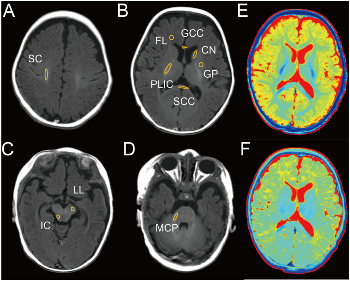
Figure 1.
Representative image of male infant brain at 3 months with delineation of ROIs on T1 image. (A) Semioval center (SC). (B) Frontal lobe (FL), posterior limb of the internal capsule (PLIC), genu of the corpus callosum (GCC), splenium of the corpus callosum (SCC), caudate nucleus (CN), globus pallidus (GP). (C) Inferior colliculus (IC), lateral lemniscus (LL). (D) Middle cerebellar peduncle (MCP). (E) T1 map. (F) T2 map.

