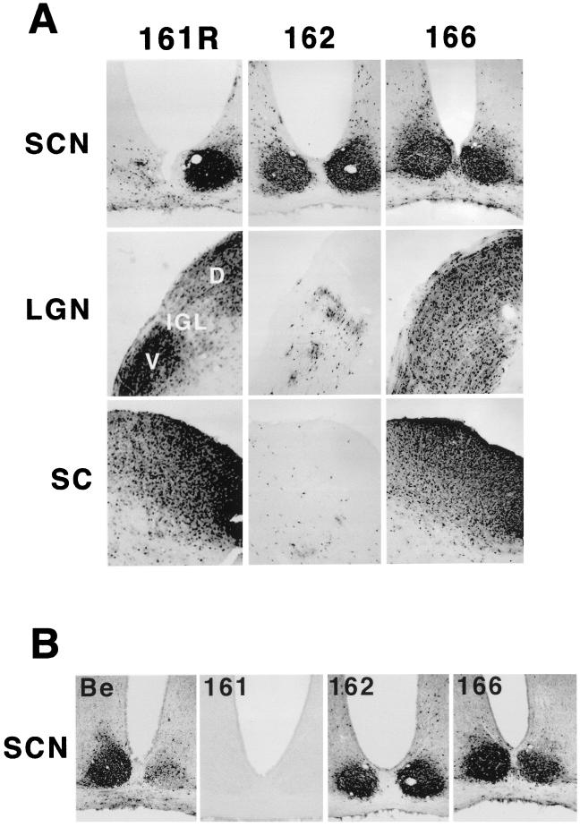
FIG. 4.
Anterograde spread of Us9 trafficking mutants in the rodent visual system. Approximately 1 × 10⁶ PFU of PRV 162 (del 46–55) and 6.8 × 10⁵ PFU of PRV 166 (L30–31A) was injected into the vitreous humor of Sprague-Dawley male rats. (A) Thirty-five-micrometer-thick coronal sections of the infected brains were examined for total PRV antigen with polyvalent antiserum Rb133, which recognizes all of the major envelope glycoproteins. (B) SCNs of PRV-infected animals stained for the Us9 protein with a polyvalent Us9-specific antiserum. Representative sections are shown for each virus. The data for the wild-type strain PRV Be, the Us9 null virus PRV 161, and the revertant virus PRV 161R have been reported previously (7) and are included here only for comparison. Due to sectioning of the PRV 161R-infected brain at an oblique angle, the image in this figure shows viral antigen in only one SCN. D, dorsal, V, ventral.

