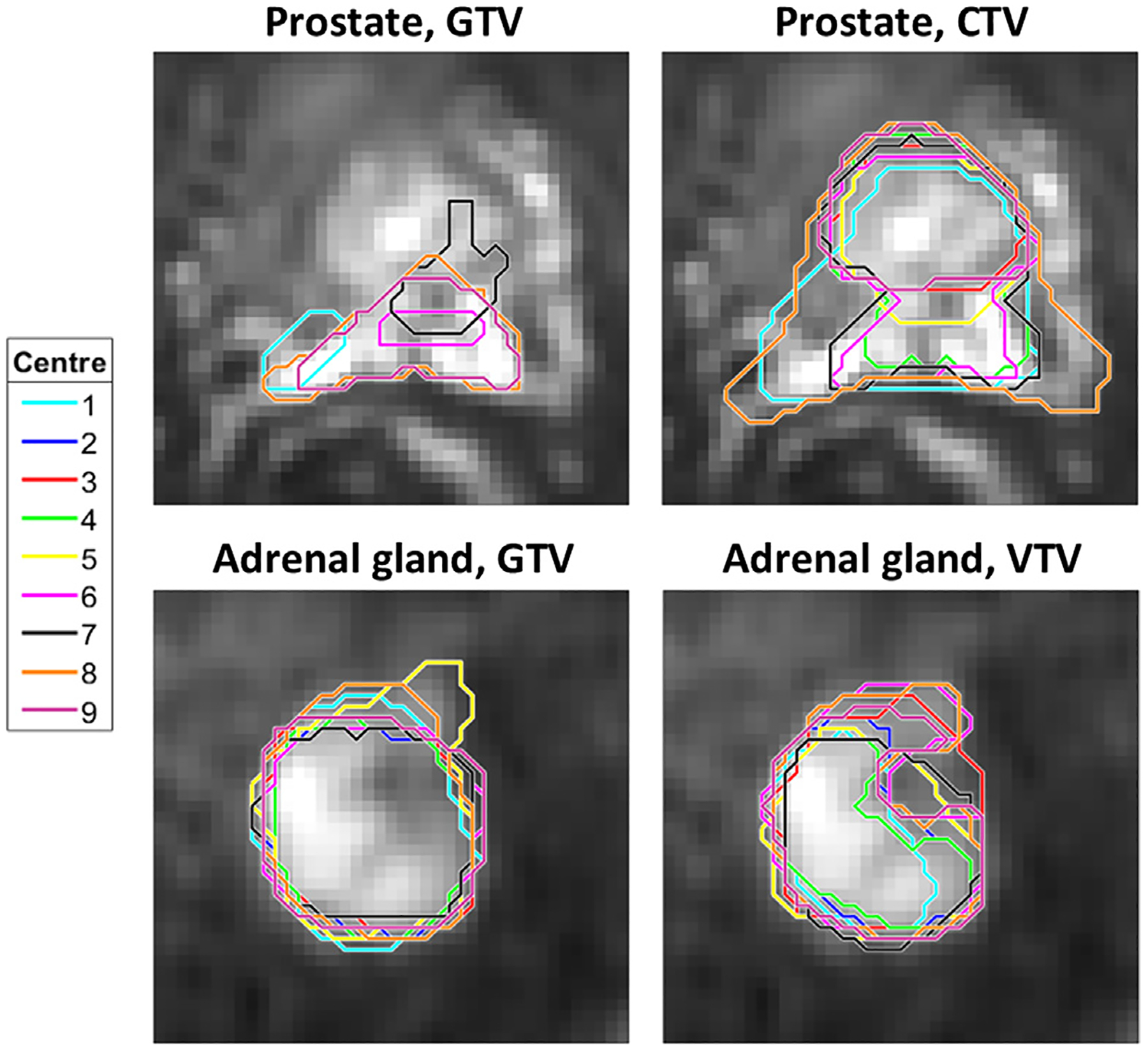
Fig. 2.

Examples of delineations. Delineations made by the nine participating centres for prostate and adrenal gland, shown on b = 500 /mm2 DWI images, cropped to an area of 7.7 × 7.7 cm2 (prostate) and 4.9 × 4.9 cm2 (adrenal gland) around the tumour. For the prostate, not all delineated contours included the shown slice, thus, only five contours are visible.
