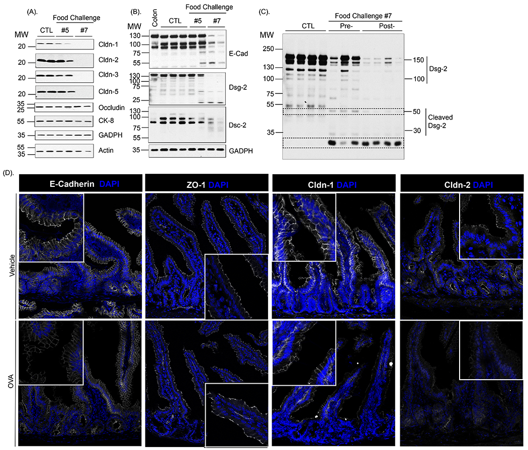
Fig. 3. Oral antigen-induced SI para-cellular dysfunction is associated with degradation of adherence and tight junction proteins.

Assessment of intestinal junctional proteins expression: Western protein analysis of (A) Claudin-1, Claudin-2, Claudin-3, Claudin-5, Occludin, Keratin-8, GADPH and Actin and (B) E-cadherin, Dsg-2, Dsc-2 and GADPH in isolated jejunal epithelial cells from untreated (CTL) and OVA-sensitized mice 30 min following the 5th and 7th oral challenge. (C) Western protein analysis of Dsg-2 in isolated jejunal epithelial cells from untreated (CTL) and OVA-sensitized mice prior to (Pre-) and 30 min following the 7th oral challenge (Post-) (A-C). Actin and GAPDH were used as a loading control. Colonic tissue was used as a positive control. MW, Molecular weight. Each column represents a single mouse (D) Immunofluorescence analysis of E-cadherin, ZO-1, Claudin-1 and Claudin-2 (white) in jejunum segments from Vehicle- and OVA-treated BALB/c WT mice within 30 min of the 7th OVA challenge. Nuclei are visualized with DAPI (blue).; (D). Representative photomicrographs of n = 5 - 7 mice per group and 5 serial SI sections per mouse.
