
Fig. 4. Oral antigen exposure leads to temporal loss of epithelial transcellular and paracellular dysfunction.
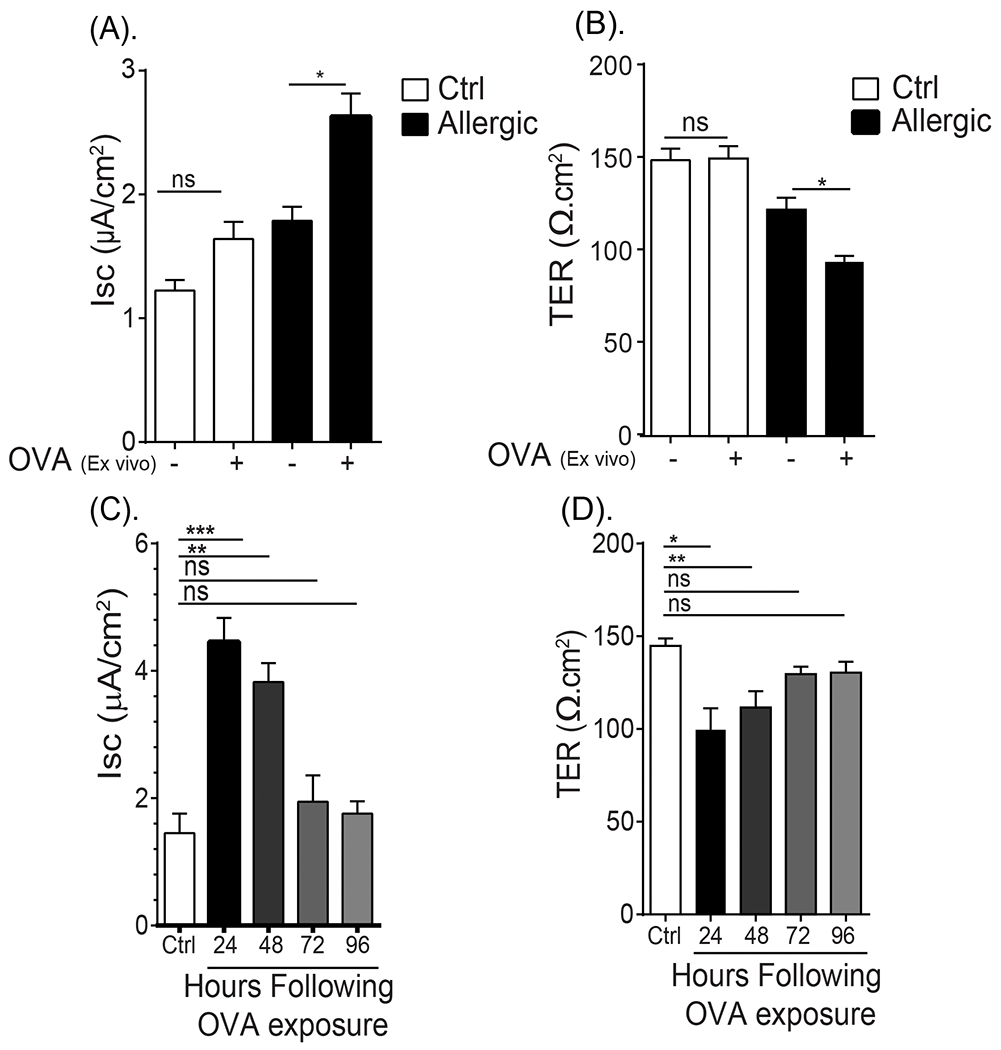
(A) Isc baseline and (B) TER of ex vivo jejunal segments from unsensitized (naïve) and OVA-sensitized and oral challenged mice (allergic) apically exposed to vehicle (PBS) or OVA in Ussing Chamber system. (C) Isc and (D) TER of ex vivo jejunal segments from unsensitized (Ctrl) and OVA-sensitized and oral challenged mice 24 - 96 hours following the seventh oral challenge. (A and B) Jejunum was removed from unsensitized (naïve) and OVA-sensitized and oral challenged mice (allergic- following 6th oral challenge) and mounted in a Ussing Chamber system and exposed to either PBS (−) or 1% OVA on the apical side and Isc and TER were recorded as described in the material and methods section. (C and D) Jejunum from unsensitized (Ctrl) and OVA-sensitized and oral challenged mice were removed 24, 48, 72 and 96 hours following seventh challenge and mounted in a Ussing Chamber system and Isc and TER were recorded as described in the material and methods section. Data are represented as the mean ± SD, n = 3 mice per group. **** P < 0.0001, *** P < 0.001, ** P < 0.01, * P < 0.05, ns > 0.05.