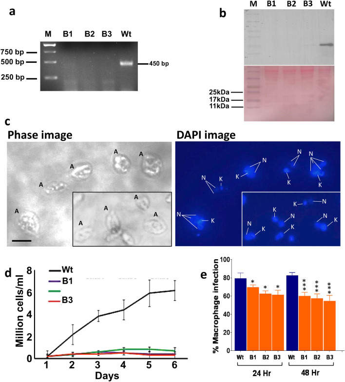
Figure 2.
A Genetic confirmation and growth characteristics of cGLP amastigotes of the 3 batches. (a) PCR confirmation of the cGLP cells to know the cells were LdCen1−/− comparing with the wild type cells (see the absence of amplification of centrin genes in the lanes from the DNA of cGLP of all the 3 batches (B1-B3). (b) Western blot analysis confirming of the cGLP cells to know the cells were LdCen1−/− comparing with the wild type cells (see the absence of centrin1 protein in lanes B1-B3 of proteins of cGLP batches B1-B3; bottom panel shows the ponceau staining of the same gel as loadings of proteins). (c) Microscopic view of the batch 1 (B1) LdCen1−/− cGLP cells to see the LdCen1−/−’s typical multinucleated (N) nature of amastigote (A) cells (with still single kinetoplast (K)) in them confirming the arrested cell division in those cells. Scale bar in all: 5 µM. (d) Growth of the three batches of the cGLP cells of LdCen1−/− axenic amastigotes displaying the growth arrested nature of the LdCen1−/− cells of all the batches as observed previously by us with the laboratory grade cells. (e) Percentage of infected human macrophages with the 3 batch parasites of LdCen1−/− in vitro at various time points 48 h post-infection. T-test: *P < 0.05, ***P < 0.0005.

