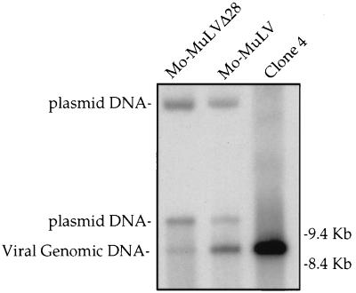
FIG. 4.
Southern blot analysis of unintegrated viral DNA synthesized after acute infection. COS-7 cells were transfected with plasmids expressing either Mo-MuLV or Mo-MuLVΔ28, and 2 days later culture supernatants were collected, normalized to contain equal amounts of RT activity, and used to infect Rat2-2 cells. Parallel cultures were also infected with wt virus from chronically infected NIH 3T3 cells (clone 4) to distinguish the reverse-transcribed linear DNA product from the transfected DNA plasmid. Low-molecular-weight DNA was isolated 18 h after infection with the indicated viruses and analyzed by Southern blot. The positions of migration of the linear viral genomic DNA and two forms of the contaminating plasmid DNA are indicated on the left, and those of the DNA markers are shown on the right.

