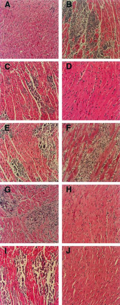
FIG. 2.
Representative histology of murine myocardium following inoculation of CVB3 and chimeric viruses. Juvenile male C3H/HeJ mice were inoculated with virus, and at 10-dpi hearts were recovered, sectioned, and stained as described in Materials and Methods. Shown are murine myocardium of negative control (A), typical myocarditis seen following inoculation of positive control CVB3/20 (B), murine myocardium following inoculation of CVB3/AS (C) and CVB3/CO (D), intratypic capsid chimeras ASP1/20 (E) and COP1/20 (F), intratypic 5′NTR chimeras AS5′/20 (G) and CO5′/20 (H), and intratypic 5′NTR/capsid chimeras AS5′P1/20 (I) and CO5′P1/20 (J). Imaged at ×200 magnification.

