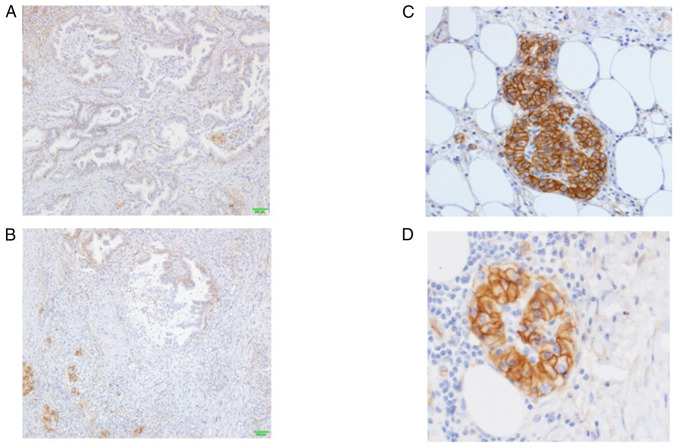
Figure 5.
Results of hENT1 immunostaining. (A) hENT1 staining showedlow-frequency and weak positivity in the central area, (B) whereas it exhibited strong staining in the invasive front of the cancer. (C) Staining of almost all cellmembranes was found in the invasive front of the cancer. (D) In the other invasive front cancer cells, strong staining of the cell membrane was found.

