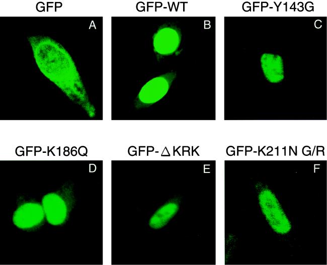
FIG. 7.
Confocal microscopic analysis of GFP-IN fusion proteins. HeLa cells were transfected with plasmid expressing GFP only (A), GFP fused to full-length WT HIV-1 IN (B), or GFP fused to the IN carrying the mutation Y143G (C), K186Q (D), ΔKRK (E), or K211N G/R (F) by using Effectene Transfection Reagent (Qiagen). At 24 h posttransfection, cells were fixed and examined with a confocal fluorescent microscope.

