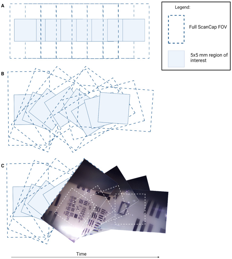
Figure 3.
Diagram of image sequence acquisition. (A) Hypothetical image capture sequence if the mirror and the camera were rotated together. (B) Actual ScanCap image sequence with rotating mirror and stationary camera. (C) Image sequence taken with ScanCap with motor rotating the mirror. In each diagram, the full ScanCap field of view (FOV) (12 × 7 mm2) and the smaller region of interest (5 × 5 mm2) are depicted. The full ScanCap FOV is utilized for mosaicking purposes and enables full coverage of the center 5 × 5 mm2 region of interest.

