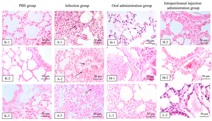
Figure 8.
Histopathological analysis in mice in the PBS group (K–1, K–2 and K–3), infected group (A–1, A–2 and A–3), oral (the H–1, M–1, and L–1 were 25.48, 12.74, and 6.37 mg/kg b.w., respectively) and intraperitoneal administration 48 h after treatment with halicin (the H–2, M–2, and L–2 were 3.69, 1.47, and 0.74 mg/kg b.w., respectively) evaluated by H&E staining (100×, scale bar: 50 μm). Note: the arrows in the images indicate varying degrees of pathological changes (pulmonary congestion, lung hemorrhage, and inflammatory cell infiltration) in the lung tissue after infection with APP S6 in mice.

