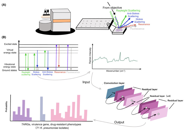
Figure 2.
Schematic overview of confocal Raman microscopy techniques, from sample preparation to spectral analysis and construction of the ResNet taxonomic model [37]. (A) illustrates the experimental setup with a confocal Raman microscope analyzing a bacterial sample, showing the various types of scattering and fluorescence phenomena that can be observed, while (B) presents a diagram of the vibrational energy levels and electronic states involved in Raman scattering phenomena, with a typical Raman spectrum resulting. Using a one-dimensional residual network with 25 total convolutional layers, Raman spectra are analyzed to predict the existence of ARGs and virulence genes or drug-resistant phenotypes.

