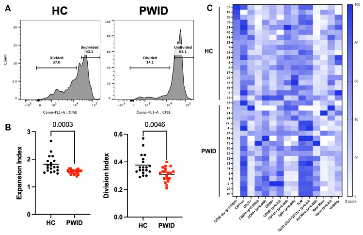
Figure 3.
Inhibition of B cell proliferation by PWID plasma. CFSE-labeled PBMC from a healthy control was cultured in IL-2, CD40L, and α-IgM for 4 d in the presence of HC (n = 19) or PWID (n = 19) plasma (1:50) samples and analyzed by flow cytometry. (A) Representative histograms gated on live CD19+ B cells. (B) Metrics of B cell proliferation. Each symbol represents an individual. (C) Frequency of divided B cells (CFSE div) and divided B cells expressing indicated marker or subset; naïve (CD21+CD27-), activated memory (CD21-CD27+), TLM (CD21-CD27-) and resting memory (CD21+CD27+) normalized to lowest frequency for each parameter. Significance determined by t-test.

