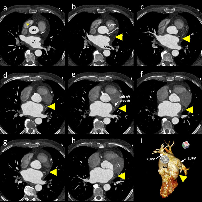
Fig. 2.
Cardiac contrast-enhanced computed tomography (CCT) images. A–H CCT axial-plane images cranial to caudal. Arrowheads show absence of the left atrial appendage in its normal position (arising from the anterolateral wall of the left atrium [LA] at the level of the left upper pulmonary vein [LUPV] and typically extending anteriorly, overlapping the left border of the pulmonary trunk and the circumflex artery). Asterisks represent the normally developed right atrial appendage. I Three-dimensional volume-rendered reconstruction of a left chamber comprehensively demonstrates the absence of the left atrial appendage (arrowhead). Ao, aorta; RVOT, right ventricular outflow tract; AV, atrioventricular; LV, left ventricle; RUPV, right upper pulmonary vein

